
What is the Connectome?
Introduction
What is the connectome?
The human connectome project
The historical backdrop to the HCP
Recent advances in connectomics
References
Further reading
The connectome is a structural map of the brain. Our current understanding of this complex and highly dynamic neuronal network is the result of a grand transatlantic collaborative venture known as the Human Connectome Project (HCP), first initiated in 2010. Since then, our knowledge about the structure of the brain and the way it functions has increased, though there is still a long way to go toward the mapping of a nanoscale connectome of a whole human brain.
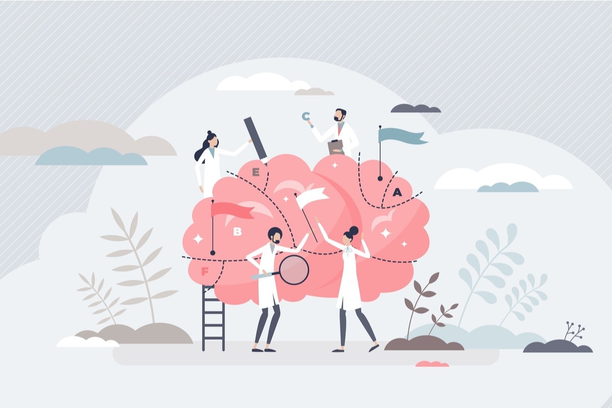 Image Credit: VectorMine/Shutterstock
Although mapping the connectome at the nanoscale level of resolution is hotly contested by some neuroscientists, understanding the brain at this level of detail could prove fruitful for progress in several fields, including artificial intelligence. The harnessing of this newfound knowledge could also prove crucial to better understanding diseases of the brain such as schizophrenia, autism, multiple sclerosis, dyslexia, Alzheimer’s, and other disorders.
What is the connectome?
The human brain is a physical object, a complex and dynamic biological network. The connectome provides comprehensive maps of structural brain connectivity to better understand the structural-functional relationship of the brain. Scientists have a long way to go until a nanoscale connectome of the whole human brain can be achieved. The brain has 1015 connections and around 100 billion neurons, so roughly as many stars as there are in the Milky Way (DeWeerdt, 2019).
Using our current-day imaging technology such a feat would take thousands of years with dozens of microscopes running all day and all night long. It is only very recently that the whole endeavor got underway, though great strides have been made since, and much progress is anticipated in the coming years.
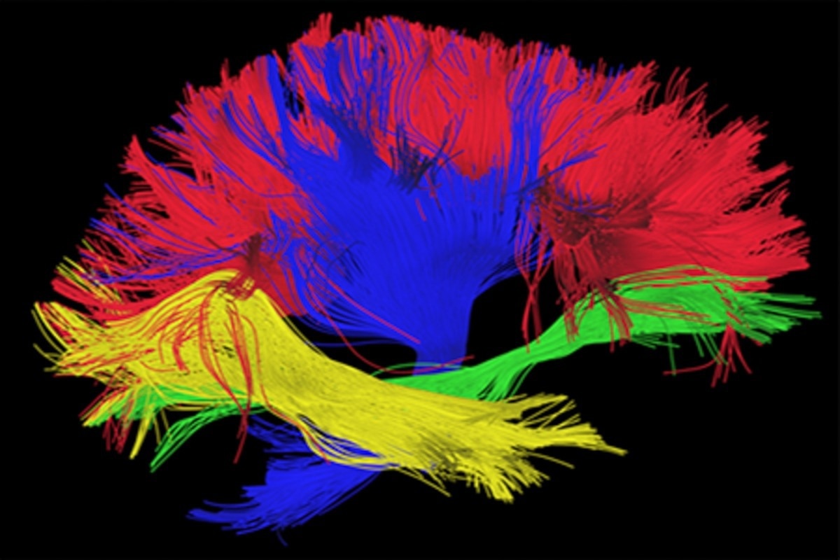 Image Credit: Image Source Trading Ltd/Shutterstock
The human connectome project
Human Connectome Project (HCP) was launched in 2010 as a venture to accelerate advances in human neuroimaging and to map structural brain connectivity. The project was met with much excitement. This was the very first time scientists had set out to map the complex wiring of the brain.
The purpose of the HCP was to provide:
- An unparalleled collection of neural data
- An interface to graphically navigate the data
- A way to know the brain better than ever before
The HCP was funded by the National Institutes of Health (NIH), who awarded a $30 million, 5-year grant to a collaborative group combing complementary strengths and comprising three centers: Washing University (WU), the University of Minnesota (UMinn), and the University of Oxford (OX). The group thus became known as the WU-Minn-OX consortium.
Read Next: Studying the Human Brain
The historical backdrop to the HCP
The foundations for the HCP lay in advances in neuroscience that occurred in the late twentieth century (Stine Elam et al., 2021).
- The emergence of Magnetic Resonance Imaging (MRI) modalities that enabled non-invasive imaging of the structure and function of the brain and investigation of connectivity using structural MRI scanning and the complementary techniques of:
- resting-state functional MRI (rfMRI)
- task-evoked functional MRI (tfMRI)
- diffusion imaging (dMRI)
- A drive to understand the ‘wiring diagram’ of the nervous system
The roots of this project can be traced back to the 1909 work of Santiago F. Ramón y Cajal.
In addition to these advances, another major milestone was the charting of synaptic connections in the nematode Caenorhabditis elegans in 1986 with advances in electron microscopy by a team led by Sydney Brenner at the University of Cambridge.
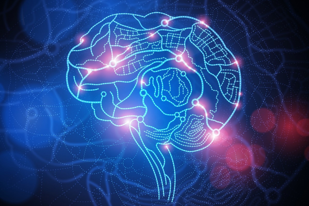 Image Credit: solarseven/Shutterstock
Recent advances in connectomics
After the synaptic mapping of Caenorhabditis elegans in the 1980s, scientists completed the task of elucidating connectome for a second species, Ciona intestinalis ––the larvae of a marine organism in 2016. Since then, the mapping of a segment of the mouse brain, the mouse-brain cubic-millimeter project, has been underway. It is no mean feat for a cubic millimeter of mouse brain comprises 100,000 neurons and the one billion connections, or synapses.
At the Allen Institute for Brain Science, based in Seattle, five transmission electron microscopes ran non-stop for five whole months. Between them, they collected over 100 million images of 25,000 slices of mouse visual cortex, each of these only 40 nanometres thick (DeWeerdt, 2019). These images were then compiled into a single 3D volume using special software developed at the institute – a project that took around three months.
The size of the dataset amounted to a whopping 2 petabytes (2 million gigabytes). To put this into perspective, it serves as a reminder that over 30 years of satellite images collected of the Earth during the Landsat missions comprised just 1.3 petabytes (DeWeerdt, 2019).
Scientists are now working on improving microscopical resolution and efficiency with a view to mapping the nanoscale connectome of the mammalian brain.
But progress does not stop there. The connectome is only able to provide details about the location of synapses, but not about their molecular composition. The next step would be the mapping of the synaptome, which could then be used in conjunction with knowledge gleaned about the connectome.
References
- DeWeerdt, S. 2019. How to Map the Brain. Nature. Doi: 10.1038/d41586-019-02208-0.
- Stine Elam, J., et al. 2021. The Human Connectome Project: A retrospective. NeuroImage. Doi: 10.1016/j.neuroimage.2021.118543.
- University of Southern California (USC) Mark and Mary Stevens Neuroimaging and Informatics Institute. n. d. Human Connectome Project. Online: http://www.humanconnectomeproject.org.
Further Reading
- All Brain Content
- Human Brain Structure
- The Impact of Climate Change on Brain Health
- Language and the Human Brain
- An Overview of Brain Development
Last Updated: Jun 24, 2022

Written by
Dr. Nicola Williams
I’m currently working as a post-doctoral fellow in the History of Science at the Leeds and Humanities Research Institute (LAHRI), at the University of Leeds. Broadly speaking my research area falls within the remit of the history of biology and history of technology in the twentieth century. More specifically I have specialist knowledge in the areas of electron microscopy and cellular and molecular biology, women in science and visual culture.
Source: Read Full Article


