
What Causes Trichomoniasis?
Trichomoniasis represents a common sexually transmitted infection that is caused by the flagellated parasitic protozoan agent Trichomonas vaginalis. Symptoms include inflammatory vaginitis in women and urethritis in men, albeit asymptomatic cases are not rare.
Nevertheless, this condition has been linked to pre-term deliveries and increased infant mortality, as well as facilitated transmission of human immunodeficiency virus (HIV).
Trichomonas vaginalis was initially discovered by a physician, Alfred Francois Donné, in 1836 who observed motile microorganisms in the purulent vaginal discharge of infected women, while a clinical entity known as trichomoniasis was first described in 1916 by Hohne. However, despite these early breakthroughs, the research on this organism was not fully launched until the 20th century.
Morphology of the causative agent
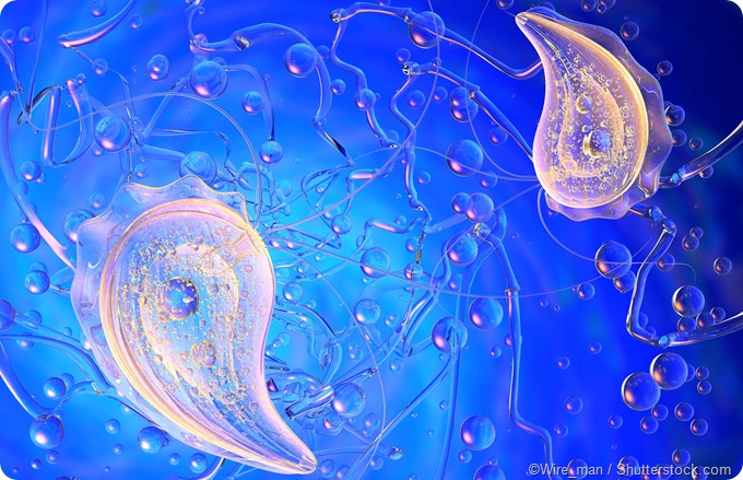
Trichomonas vaginalis is a parasitic protozoan that, akin to some other protozoan parasites, exists only as a trophozoite and does not form a cyst. This human pathogen may be variable in size and shape, but always carries four anterior flagella and a recurrent fifth one that is attached to the parasite body by an undulating membrane. This results in a distinctive quivering motility.
A hyaline slender rod-like structure, known as axostyle, longitudinally bisects this protozoan. It protrudes through the posterior part of the parasite, terminating in a sharp point. This structure is quite important, as it is thought to anchor Trichomonas vaginalis to the vaginal epithelial cells.
This parasite also has hydrogenosomes, which are electron-dense spherical organelles or granules with a diameter of 0.5 micrometers. Biochemical studies have demonstrated that these organelles are functionally very similar to mitochondria, as they harbor enzymes of the tricarboxylic acid cycle used to synthesize adenosine triphosphates.
Growth and life cycle of Trichomonas vaginalis
Scanning electron microscopy processing has clearly shown that Trichomonas vaginalis is most often ovoid in shape, between 6 to 28 millimeters long and 5 to 12 millimeters wide. An optimal condition for its in vitro growth is a pH between 6.0 and 6.3, although a wide range of pH values may be suitable for trichomonads.
This flagellate reproduces by longitudinal binary fission, whilst preserving the nuclear membrane in the process. Several oversized round forms of Trichomonas vaginalis are described in dividing growth phase culture – these are, most notably, those without flagella, those with flagella and dividing nucleus, as well as those with flagella and manifold nuclei. The prevailing school of thought is that these forms are not stages in the life cycle; rather, these arise during certain disadvantageous conditions.
Genetic and clinical intricacies of the organism
The genome sequence of Trichomonas vaginalis was published in 2007, which prompted other studies that revealed a two-type population structure in a worldwide sample of affected women. A recent study has shown that those diagnosed with this parasite were infected with either type 1 or type 2 Trichomonas in near-equal percentages, while 10 percent of women carried both types.
Isolates of Trichomonas vaginalis that were type 1 were easier to detect with commonly employed diagnostic procedures (such as wet mount) and may signify symptomatic individuals with high parasite loads. On the other hand, those that were type 2 necessitated higher concentrations of drugs to achieve lethal effect on the parasite; hence they may be linked to antibiotic resistance.
An interesting factoid is a double-stranded RNA virus (commonly dubbed Trichomonas vaginalis virus) that is detected in 73 percent of type 1 Trichomonas isolates, but only in 2.5 percent of type 2 Trichomonas isolates. It is thought that the presence of this virus can modulate the pathogenicity of the parasite by altering the expression of cysteine proteinases and surface molecules.
Sources
- http://www.antimicrobe.org/new/b65.asp
- http://jcm.asm.org/content/54/1/7.long
- https://www.ncbi.nlm.nih.gov/pmc/articles/PMC3586271/
- www.scielo.cl/scielo.php
- www.academicjournals.org/…/59BB4D314234
- Strelkauskas A, Edwards A, Fahnert B, Pryor G, Strelkauskas J. Microbiology: A Clinical Approach, Second Edition. Garland Science, Taylor & Francis Group, 2016; pp. 328-329.
Further Reading
- All Sexually Transmitted Disease (STD) Content
- What is an STD (Sexually Transmitted Disease)?
- STD Pathophysiology
- STD Prevention and Treatment
- STD Diagnosis
Last Updated: Feb 27, 2019

Written by
Dr. Tomislav Meštrović
Dr. Tomislav Meštrović is a medical doctor (MD) with a Ph.D. in biomedical and health sciences, specialist in the field of clinical microbiology, and an Assistant Professor at Croatia's youngest university – University North. In addition to his interest in clinical, research and lecturing activities, his immense passion for medical writing and scientific communication goes back to his student days. He enjoys contributing back to the community. In his spare time, Tomislav is a movie buff and an avid traveler.
Source: Read Full Article


