
What is Immunofluorescent Array Tomography?
Immunofluorescent array tomography is an imaging technique that relies on fluorescent staining to generate high-resolution images. While it was originally developed for the study of neurons, its applications within a range of research areas have rapidly grown, and it is now an established method of studying aortic aneurysms, synapses, neurons, organelles, and antibodies.
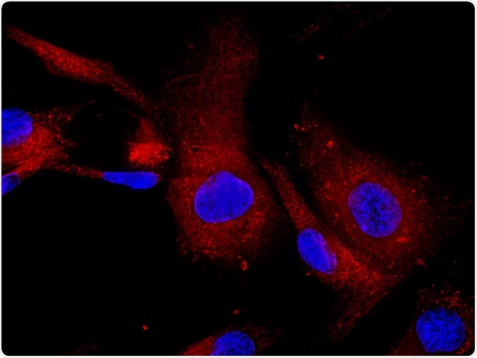 Image Credit: Vshivkova / Shutterstock.com
With the equipment available and the technique having become increasingly sophisticated, we can expect applications of this method to continue to grow.
What is immunofluorescent array tomography and how does it work
Originally developed for applications in neuroscience, immunofluorescent array tomography is a three-dimensional microscopy method that generates reliable high-resolution images of substrates.
The method was initially developed by Kristina D. Micheva and Stephen J. Smith at the University of Stanford, and it works by imaging two-dimensional arrays of ultra-thin specimens of solid substrates. First, the tissue to be studied is embedded within resin and sliced into ultra-thin sections that are then adhered to microscope slides. These arrays are stained with fluorescent antibodies or other fluorescent stains and eluted and restrained numerous times, to create ultra-high-resolution 3D images.
Fluorophores are a kind of fluorescent chemical compound that can absorb and re-emit light at two distinct wavelengths. They are used as the staining agents in this process. When exposed to light of a suitable wavelength the fluorophores fluoresce: the light is absorbed by the fluorescent molecules, which excites constituent electrons causing them to rise to a higher energy level.
These electrons only remain in this excited state for a brief time, and as they return to their ground state they release the same quanta of energy as was absorbed in the form of photons. These emitted photons are what are detected during immunofluorescent array tomography: the emitted light is registered by either a fluorescent microscope or a confocal microscope, which is what produces the detailed, ultra-high-resolution volumetric images of the substrates.
The past two decades have seen biological sciences increasingly use fluorescence methods for various research purposes. Due to this, these methods have grown in sophistication, resulting in current methodologies and availably equipment for immunofluorescent array tomography having reached an advanced level.
Immunofluorescent array tomography in research
While it was first developed with the aim to use it to look at and study neural circuits, immunofluorescent array tomography has already proven its functionality in studying various fields of biological science, not limited to its original intended field.
Neural circuits
The exact intricate biological functions of systems within the brain have often eluded the exploration of previous imaging techniques. This is because these techniques have failed to provide the fine-grain details of the molecular architecture of targeted tissue, which has led to the intricate circuits and synaptic architectures failing to be seen in high definition.
The technique of immunofluorescent array tomography offers advantages to the study of neural circuits in that it provides high definition imagery, offering a unique look into the features of brain molecular architecture.
Organelles
Organelles are structures that exist within a cell’s intracellular membrane. The family of organelles includes structures such as mitochondria, ribosomes, the endoplasmic reticulum, and the nucleus. Numerous studies have shown that immunofluorescent array tomography is successful in providing high definition and reliable imagery of the structure of organelles. This information has proved to be invaluable in the role of organelle function in numerous pathologies and diseases.
Antibody characterization
To be able to use antibodies effectively in different applications, their function and behavior in relation to specific synaptic molecules must be validated. Immunofluorescent array tomography has been used successfully in numerous studies that have sought to characterize antibodies.
It works through using immunofluorescent labeling to detect whether the puncta from a candidate antibody belong to a certain synapse. The method is now considered a robust and efficient tool for antibody characterization. It provides a method for comparing numerous antibodies against the same target synapse, helping to push forward the knowledge base for immunotherapy research.
Synapse analysis
The study of neurodegenerative disease, developmental disease, and psychiatric illness benefit greatly from the postmortem study of synapses. Previous methods of imaging, such as light microscopy and electron microscopy, had the respective limitations of low axial resolution and difficulty in preserving and analyzing ultrastructure.
Immunofluorescent array tomography addresses these problems, and solves them by embedding ultra-thin sections of autopsy tissue in resin. Imaging of these sections results in a resolution able to see tens of thousands of synapses.
Study of aortic aneurysms
The study of aortic aneurysm development has benefited from immunofluorescent array tomography. While the elastase-perfusion AAA murine model, and other aneurysm animal models have previously been relied upon to gain an understanding of the disease, immunofluorescent array tomography has offered a technique that gives a view of the finer details of the microarchitecture and cellular morphology during abdominal aortic aneurysm.
Future directions
Immunofluorescent array tomography has been shown to a useful technique in many areas of research for generating reliable, high-resolution images. It is becoming increasingly relied upon in biological research as it has been found to offer benefits over previously relied upon methods.
It has already developed greatly in its level of sophistication, and it is expected to find further uses in the research and exploration of biological mechanisms, aiding in developing insights into disease and potential therapies.
Sources:
- Docs.abcam.com. (2019). [online] Available at: https://docs.abcam.com/pdf/protocols/array-tomography-full-protocol.pdf [Accessed 22 Oct. 2019].
- Docs.abcam.com. (2019). [online] Available at: https://docs.abcam.com/pdf/protocols/array-tomography-protocol-immunostaining-elution-and-data-analysis.pdf [Accessed 22 Oct. 2019].
- Kay, K., Smith, C., Wright, A., Serrano-Pozo, A., Pooler, A., Koffie, R., Bastin, M., Bak, T., Abrahams, S., Kopeikina, K., McGuone, D., Frosch, M., Gillingwater, T., Hyman, B. and Spires-Jones, T. (2013). Studying synapses in human brain with array tomography and electron microscopy. Nature Protocols, 8(7), pp.1366-1380. https://www.nature.com/articles/nprot.2013.078
- Micheva, K. and Smith, S. (2007). Array Tomography: A New Tool for Imaging the Molecular Architecture and Ultrastructure of Neural Circuits. Neuron, 55(1), pp.25-36. https://www.ncbi.nlm.nih.gov/pubmed/17610815
- Saatchi, S., Azuma, J., Wanchoo, N., Smith, S., Yock, P., Taylor, C. and Tsao, P. (2011). Three-Dimensional Microstructural Changes in Murine Abdominal Aortic Aneurysms Quantified Using Immunofluorescent Array Tomography. Journal of Histochemistry & Cytochemistry, 60(2), pp.97-109. https://journals.sagepub.com/doi/full/10.1369/0022155411433066
- Simhal, A., Gong, B., Trimmer, J., Weinberg, R., Smith, S., Sapiro, G. and Micheva, K. (2018). A Computational Synaptic Antibody Characterization Tool for Array Tomography. Frontiers in Neuroanatomy, 12. https://www.ncbi.nlm.nih.gov/pubmed/30065633
Further Reading
- All Fluorescence Content
- What is Fluorescence Spectroscopy?
- How do Epifluorescence Microscopes Work?
- GFP-tagging in Fluorescence Microscopy
- Fluorescence Quenching
Last Updated: Dec 10, 2019

Written by
Sarah Moore
After studying Psychology and then Neuroscience, Sarah quickly found her enjoyment for researching and writing research papers; turning to a passion to connect ideas with people through writing.
Source: Read Full Article


