Study shows mink can be infected by Omicron and efficiently transmit the virus to other mink
Coronavirus disease 2019 (COVID-19) has been widely believed to be a zoonotic disease. While the theory that the disease jumped from a bat into a human gained by far the most media attention, many scientists believe that the disease likely moved into more than one alternate host before infecting humans. Since the spread of the disease around the globe, it has been found that several other species are susceptible to infection with severe acute respiratory syndrome coronavirus 2 (SARS-CoV-2), notably including felines and mink. Researchers have been examining the pathology and spread of the Omicron variant in mink.
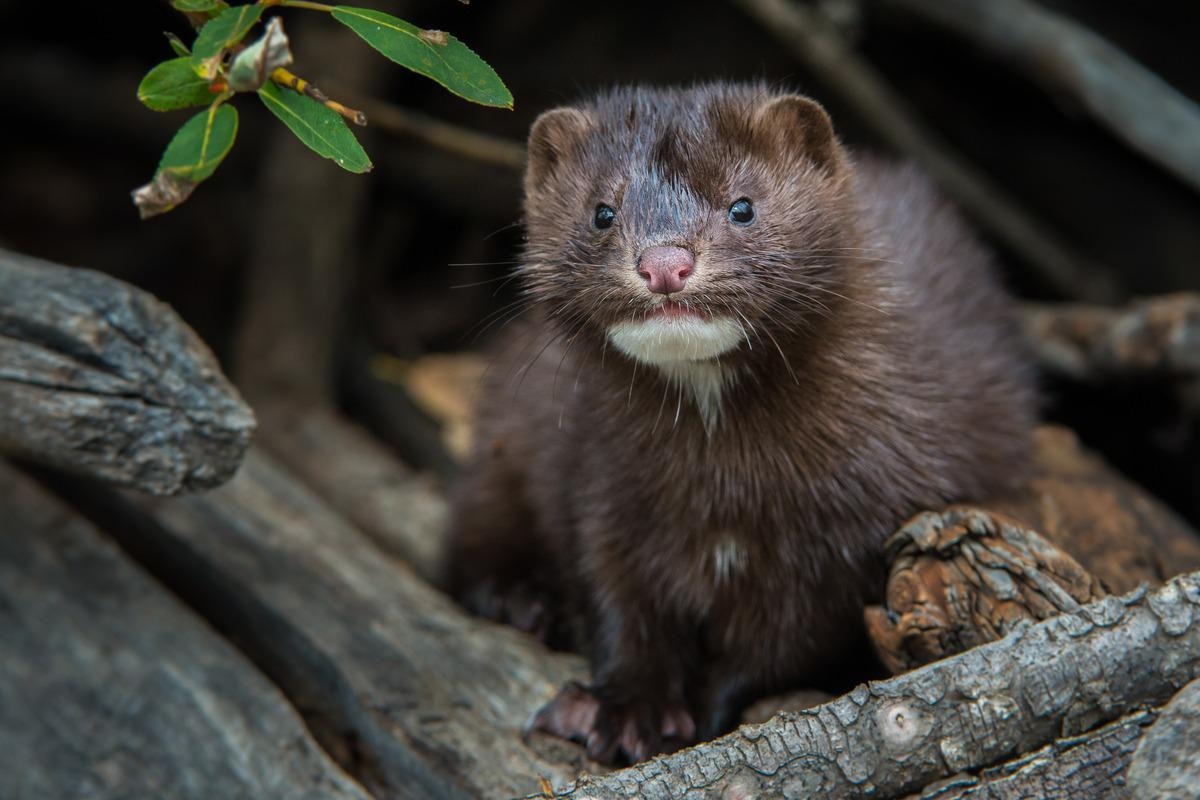 Study: Experimental infection of mink with SARS-COV-2 Omicron (BA.1) variant leads to symptomatic disease with lung pathology and transmission. Image Credit: Ghost Bear/Shutterstock
A preprint version of the study is available on the bioRxiv* server while the article undergoes peer review.
The study
The researchers infected three male minks with 4e5 PFU of the omicron variant intranasally. The minks were then followed for seven days, with daily saliva samples and histopathological evaluations of the upper and lower respiratory tracts on the final day of the follow-up. All of the mink were successfully infected, and none showed signs of severe disease within the seven days. The scientists observed a range of symptoms, including lethargy, anorexia, diarrhea, nasal and lacrimal discharge, and sneezing. Saliva samples showed positive PCR tests after a single day and remained positive for the rest of the experiment.
During this period, the scientists decided to test if minks could transmit the Omicron variant. To accomplish this, they placed two uninfected minks in separate cages nearby the cages of the infected mink (10 to 20cm away). Both uninfected minks developed symptoms rapidly and showed consistent positive PCR test results from day three onwards.
When examining the respiratory tracts, they found hyperemia of respiratory mucosa with viscous exudate and non-collapsed, wet pulmonary lobes. All the mink showed these changes. In the nose, multifocal degeneration was seen alongside the loss of respiratory epithelium, and both mucosal and submucosal neutrophilic infiltration could be seen. In the lumen, the researchers found sloughed epithelial cells, mucinous material, degenerated neutrophils, and viral nucleoproteins could be found distributed throughout the respiratory system, within sloughed cells and the mucosal respiratory epithelium.
The scientists found that the olfactory epithelium was inconsistent and only mildly affected with focal viral antigen detection. The lungs showed clear pathology. Both the recipient minks and two inoculated ones showed pulmonary lesions associated with viral antigen expression and characterized by multifocal to coalescing alveolar damage with degeneration and/or necrosis of alveolar septa, infrequent hyalin membrane formation, and variable proliferation of type II pneumocytes.
Alveolar spaces contained macrophages alongside sloughed cells, edema, and hemorrhage, and the researchers could observe bronchiolar epithelial degeneration and hyperplasia in some but not all. The lumen here was filled with sloughed cells and neutrophils. Bronchi were found to be lined by hyperplastic epithelium with a greater than the usual number of goblet cells. Researchers found evidence of vasculitis, perivasculitis, perivasculitis, perivascular, and peri-bronchial edema in most infected mink.
In one particular mink, the researchers found significantly thickened alveolar septa by mononuclear cells, marked proliferation of type II pneumocytes, intra-alveolar macrophages, few syncytial cells, bronchi, and bronchiolar epithelial cell hyperplasia, vasculitis, and perivasculitis. Strangely, they could not detect viral antigen in this mink at all, and no mink showed detectable viral antigen in the epithelium of bronchi or bronchioles.
The conclusion
The authors acknowledge that while some of the clinical signs could have arisen due to stress from the change of environments, the positive PCR tests and the symptoms in line with what has been previously observed in other infections show that Omicron can cause symptomatic infection in mink. They have also proven that mink-to-mink transmission is possible.
They claim that while there is no evidence of mink-to-human transmission, it seems likely based on their results and information from other variants. Information from other variants notwithstanding, nothing in this exploration has shown any significant reason to assume this.
However, the scientists have increased our understanding of the pathological implications of Omicron infections in mink. The research they have performed should help inform those involved in mink farming. A better understanding of the symptoms present in infected mink could help control outbreaks before they become overwhelming.
*Important notice
bioRxiv publishes preliminary scientific reports that are not peer-reviewed and, therefore, should not be regarded as conclusive, guide clinical practice/health-related behavior, or treated as established information.
-
Virtanen, J. et al. (2022) "Experimental infection of mink with SARS-COV-2 Omicron (BA.1) variant leads to symptomatic disease with lung pathology and transmission". bioRxiv. doi: 10.1101/2022.02.16.480524. https://www.biorxiv.org/content/10.1101/2022.02.16.480524v1
Posted in: Medical Science News | Medical Research News | Disease/Infection News
Tags: Anorexia, Antigen, Bronchi, Cell, Coronavirus, Coronavirus Disease COVID-19, covid-19, Diarrhea, Edema, Hyperplasia, Lethargy, Lungs, Membrane, Necrosis, Neutrophils, Omicron, Pathology, Proliferation, Research, Respiratory, SARS, SARS-CoV-2, Severe Acute Respiratory, Severe Acute Respiratory Syndrome, Sneezing, Stress, Syndrome, Vasculitis, Virus

Written by
Sam Hancock
Sam completed his MSci in Genetics at the University of Nottingham in 2019, fuelled initially by an interest in genetic ageing. As part of his degree, he also investigated the role of rnh genes in originless replication in archaea.
Source: Read Full Article