Study indicates that macrophages that reside in the lymph nodes contribute to the initial Zika virus spread
In a recent study published in Cell Reports, researchers mapped Zika virus infections in the lymph nodes to determine how the Zika virus enters from different points in the skin and disseminates through the body.
Background
Immunology eBook

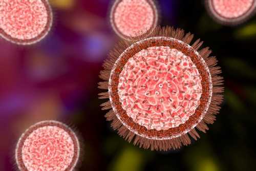
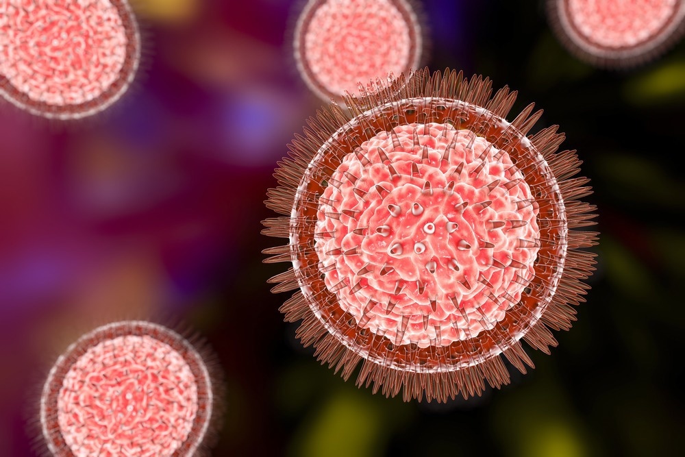
Zika virus is a positive-sense, single-stranded ribonucleic acid (RNA) virus belonging to the Flavivirus genus transmitted by mosquitoes. While most Zika virus infections have been mild, with some patients exhibiting prolonged viremia cases, recent studies have reported microcephaly and congenital neurodevelopmental disease associated with Zika virus infections. This has accelerated the research on the pathogenesis of Zika virus infections, therapeutic options, and candidate vaccines. However, no approved therapy or vaccine currently exists against the Zika virus.
Zika virus enters the bloodstream through the skin and accesses different tissues such as the brain and the placenta, decidua, and the fetus in pregnant women. Cellular targets such as the Tyro3, Mertk, and Axl receptor tyrosine kinases have been explored to understand how the Zika virus disseminates through the skin into peripheral tissue.
Monocytes, which are abundant in blood and have access to most tissues in the body, are the primary type of cells infected by the Zika virus and have been studied to understand the movement of the virus. However, the role of the cells within the lymph nodes involved in the early movement of the virus during the dissemination process remains unclear.
About the study
In the present study, the researchers conducted footpad inoculation of the interferon alpha receptor 1 (Ifnar1–/–) mouse model with the Zika virus. The early stages of the viral delivery kinetics were observed in the popliteal lymph node (PLN), which drains in the hindfoot. The reasoning was that if skin cells were required for viral replication, high viral titers would not be detected in the PLN.
After inoculation with 104 focus-forming units of Zika virus, PLNs were harvested at multiple time points within one hour to determine the viral titers through focus-formation assays. The infectious virus titers from serum samples and PLNs were also quantified every four hours for the first 32 hours post-infection.
Five distinct macrophage populations are found in lymph nodes related to specific immune functions. Macrophages in the nodal sinuses closest to the incoming lymph vessels are called subcapsular sinus macrophages, while those in the nodal sinuses near the exiting lymph vessels are called medullary sinus macrophages.
The researchers investigated whether the macrophages in the lymphoid sinuses capture the Zika virus by imaging PLN cross-sections harvested from the inoculated mice at eight to 24 hours post-infection.
Necroptosis or pyroptosis and inflammasome activation cause attrition in the subcapsular sinus macrophages when inflammation is stimulated by dead or live viruses. This feature was used to measure the sensing of Zika virus in the lymph nodes by lymph node macrophages through flow cytometry of PLN and iliac lymph nodes harvested 72 hours post-infection.
Furthermore, Siglec1-cre mouse models with CD169+ cells expressing Cre recombinase were crossed with Ifnar1-floxed mice to conditionally delete IFN-I signaling in CD169+ macrophages and create CD169 conditional knockout mice. This mouse model was used to understand the role of lymph node macrophages in disseminating the Zika virus.
Additionally, based on the role of monocytes in disseminating the Zika virus in mice, the involvement of Ly6chigh monocytes in the early dissemination of the Zika virus was also examined. The role of CD169+ macrophages in sustained viremia in Ifnar1–/– mice five days post-infection was also examined.
Results
The results reported that migratory immune cells, such as monocytes, were not involved in the early dissemination of the Zika virus in the blood. Monocyte involvement is thought to occur during the spread of the Zika virus from blood to other tissues. Zika virus was seen to infect the resident CD169+ macrophages in the lymph nodes that released the virus into the downstream lymph nodes.
While studies have implicated dermal dendritic cells in disseminating flaviviruses such as the Zika virus from the skin, the results suggested that the Zika virus is already present in the blood before dermal dendritic cells migrate to lymph nodes in large numbers. Furthermore, infection of CD169+ macrophages was enough to initiate viremia. The use of CD169 conditional knockout mice showed that infection of a small number of lymph node CD169+ macrophages produced a detectable level of viremia 12 hours post-infection.
Conclusions
Overall, the findings suggested that the early stages of Zika virus infection and replication occur in CD169+ macrophages in the lymph nodes, leading to viremia. Furthermore, the Zika virus can disseminate to blood in the absence of monocytes or dermal dendritic cells. However, CD169+ macrophages are largely involved in the early dissemination, and infection of these cells alone does not result in morbidity.
- Reynoso, G. et al. (2023) "Zika virus spreads through infection of lymph node-resident macrophages", Cell Reports, 42(2), p. 112126. doi: 10.1016/j.celrep.2023.112126. https://www.sciencedirect.com/science/article/pii/S2211124723001377
Posted in: Medical Science News | Medical Research News | Disease/Infection News
Tags: Blood, Brain, Cell, Cytometry, Flow Cytometry, Imaging, Inflammasome, Inflammation, Interferon, Knockout, Lymph Node, Lymph Nodes, Macrophage, Microcephaly, Monocyte, Mouse Model, Necroptosis, Placenta, Receptor, Research, Ribonucleic Acid, RNA, Skin, Skin Cells, Tyrosine, Vaccine, Virus, Zika Virus
.jpg)
Written by
Dr. Chinta Sidharthan
Chinta Sidharthan is a writer based in Bangalore, India. Her academic background is in evolutionary biology and genetics, and she has extensive experience in scientific research, teaching, science writing, and herpetology. Chinta holds a Ph.D. in evolutionary biology from the Indian Institute of Science and is passionate about science education, writing, animals, wildlife, and conservation. For her doctoral research, she explored the origins and diversification of blindsnakes in India, as a part of which she did extensive fieldwork in the jungles of southern India. She has received the Canadian Governor General’s bronze medal and Bangalore University gold medal for academic excellence and published her research in high-impact journals.
Source: Read Full Article