Research examines the roles of cytokines during SARS-CoV-2 infection in rhesus macaques
In a recent study posted to the bioRxiv* preprint server, researchers in the United States assessed the role of two cytokines, interferon-gamma (IFN-γ) and interleukin-10 (IL-10), in regulating immune cell responses and inflammation early during severe acute respiratory syndrome coronavirus 2 (SARS-CoV-2) infection in rhesus macaques.
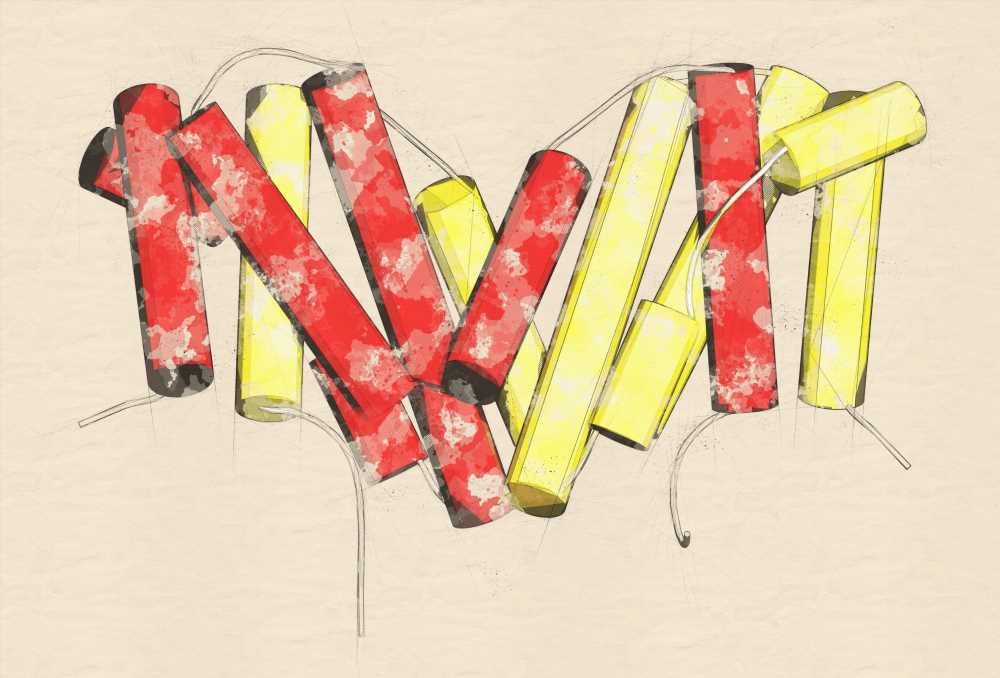
 Study: IL-10 suppresses T cell expansion while promoting tissue-resident memory cell formation during SARS-CoV-2 infection in rhesus macaques. Image Credit: StudioMolekuul / Shutterstock
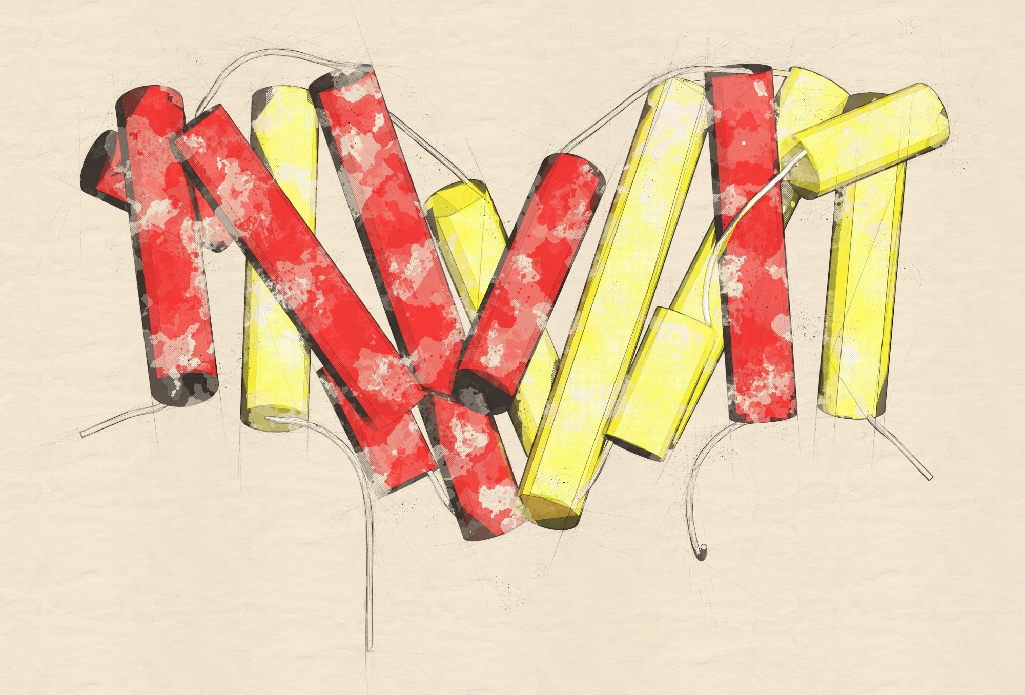
Background
While IFNγ is a pro-inflammatory cytokine, IL-10 is an anti-inflammatory cytokine. These two distinct pathways determine the balance of inflammation and SARS-CoV-2 replication at the onset of coronavirus disease 2019 (COVID-19) or during its early phases. However, studies have barely investigated the significance of these cytokines during SARS-CoV-2 infection.
About the study
In the present study, researchers used a non-human primate (NPH) model to measure changes in lung inflammation, viral replication, and cellular immune responses against SARS-CoV-2 after blocking cytokine pathways. The study had a pre-specified endpoint between days 28 and 35 post-infection.
The team created three treatment groups, each comprising five male rhesus macaques aged 2.5 to five years and weighing 3.5 to five kilograms. They infected test animals with SARS-CoV-2 in five waves, with each infection wave infecting one animal per treatment group (a total of three).
The researchers used HEK-BlueTM reporter cell lines for quantifying in vitro IL-10 and IFNγ signaling. A color change reaction indicated the magnitude of cytokine signaling, which the team quantified using a spectrophotometer at 650 nanometers. They used f-fluorodeoxyglucose (FDG)-positron emission tomography (PET)/computerized tomography (CT) to image the chest region of infected macaques. They identified volumes of interest (VOI) or lesions in each chest scan procured on day 2 or day 6, which they transferred to o the aligned PET/CT images to assess the change in lesion volume or FDG uptake.
During the necropsy of the animals, the team handled the lesions that appeared to be continuous in the PET/CT but resulted from inflammation in more than one lung lobe separately. Additionally, they scanned the upper abdomen, including the spleen and transverse colon, and the head and neck. It helped them determine (18F)FDG uptake in tonsils and nasal turbinates.
Finally, the researchers harvested the lungs and attached airways, nasal turbinates, salivary gland, tonsil, spleen, tissues, and lymph nodes of the euthanized animals. In the lungs, they assessed the parenchyma localization of T cells, whereas they used lymph node tissues for ribonucleic acid (RNA) isolation, histological analyses, and single-cell preparations for flow cytometry.
Study findings
Although IFNγ blockade reduced germinal center formation in reactive lymph nodes, it had little or no effect on any features of B or T cell responses. The researchers did not evaluate any possible impact of IFNγ on myeloid cell function, even if there were any. However, they found some interesting effects of IL-10 on SARS-CoV-2-specific T cell responses.
First, IL-10 inhibited the magnitude of virus-specific T cell responses in the circulation, lower airways, and pulmonary lymph nodes. Analyzing the expression of the marker of proliferation Ki-67 gene confirmed that IL-10 did not prolong the cycling of virus-specific T cells but mediated them during the early clonal burst. Second, IL-10 promoted the rate at which SARS-CoV2-specific T cells differentiated into tissue-resident memory T (TRM) cells at the mucosal surfaces of the lower airways, likely by triggering monocytes to increase the production of tumor necrosis factor beta (TGFβ), which, in turn, promoted TRM cells phenotype.
Surprisingly, rhesus macaques showed no SARS-CoV-2-specific T cell responses in the nasal mucosa. In fact, IL-10 blockade reduced the number of TRM cells in their nasal mucosa. However, studies in mice have detected SARS-CoV-2-specific TRM cells in the nasal mucosa. The study data pointed to the possibility that increasing IFΝγ might lead to enhanced control of SARS-CoV-2 replication. Likewise, providing exogenous IL-10 at the time of mucosal vaccination could promote the formation of tissue-resident memory T cells. In other words, the study data might have implications for targeting these cytokines as adjuvants for COVID-19 vaccination. The authors cautioned that the study results were restricted to the setting of mild COVID-19, and IFNγ and IL-10 might have different functional roles during severe COVID-19. Future studies using NHP models of COVID-19 pneumonia could shed light on cellular and molecular mechanisms of immunity-mediated lung damage.
Conclusions
The study results showed that both cytokines, IFNγ and IL-10, did not play a critical role in controlling SARS-CoV-2 replication in the rhesus macaque model. IFNγ blockade decreased lung inflammation to some extent but did not impact innate lymphocytes, neutralizing antibodies, or antigen-specific T cells. On the other hand, IL-10 blockade transiently increased lung inflammation, suppressed the accumulation of SARS-CoV-2-specific T cells in the lower airways, and promoted TRM at respiratory mucosal surfaces. Since these cytokines did not substantially affect viral load, SARS-CoV-2 infection in all the test animals eventually resolved.
*Important notice
bioRxiv publishes preliminary scientific reports that are not peer-reviewed and, therefore, should not be regarded as conclusive, guide clinical practice/health-related behavior, or treated as established information.
- IL-10 suppresses T cell expansion while promoting tissue-resident memory cell formation during SARS-CoV-2 infection in rhesus macaques, Christine E. Nelson, Taylor W. Foreman, Keith D. Kauffman, Shunsuke Sakai, Joel D. Fleegle, Felipe Gomez, NIAID/DIR Tuberculosis Imaging Program, Cyril Le Nouen, Xueqiao Liu, Tracey L. Burdette, Nicole L. Garza, Bernard A. P. Lafont, Kelsie Brooks, Cecilia S. Lindestam Arlehamn, Daniela Weiskopf, Alessandro Sette, Heather D. Hickman, Ursula J. Buchhholz, Reed F. Johnson, Jason M. Brenchley, Laura E. Via, Daniel L. Barber, bioRxiv pre-print 2022, DOI: https://doi.org/10.1101/2022.09.13.507852, https://www.biorxiv.org/content/10.1101/2022.09.13.507852v1
Posted in: Medical Science News | Medical Research News | Disease/Infection News
Tags: Antibodies, Antigen, Anti-Inflammatory, Cell, Coronavirus, Coronavirus Disease COVID-19, covid-19, CT, Cycling, Cytokine, Cytokines, Cytometry, Flow Cytometry, Fluorodeoxyglucose, Gene, immunity, in vitro, Inflammation, Interferon, Interferon-gamma, Interleukin, Lungs, Lymph Node, Lymph Nodes, Neck, Necrosis, Phenotype, Pneumonia, Positron Emission Tomography, Proliferation, Research, Respiratory, Ribonucleic Acid, RNA, Salivary Gland, SARS, SARS-CoV-2, Severe Acute Respiratory, Severe Acute Respiratory Syndrome, Spectrophotometer, Spleen, Syndrome, Tomography, Tonsil, Tumor, Tumor Necrosis Factor, Virus

Written by
Neha Mathur
Neha is a digital marketing professional based in Gurugram, India. She has a Master’s degree from the University of Rajasthan with a specialization in Biotechnology in 2008. She has experience in pre-clinical research as part of her research project in The Department of Toxicology at the prestigious Central Drug Research Institute (CDRI), Lucknow, India. She also holds a certification in C++ programming.
Source: Read Full Article