Infrared Imaging Explored for Diabetic Retinopathy Screening
NEW YORK, NY — Screening near-infrared reflectance imaging could identify cases of diabetic retinopathy for early treatment, researchers say.
In a retrospective study of near-infrared images acquired through standard optical coherence tomography (OCT) equipment, researchers were able to distinguish eyes with moderate, severe, or proliferative diabetic retinopathy from eyes without the condition.
“That boundary was useful because it separates people who need to be seen in the clinic from those who don’t,” Mina M. Naguib, MD, a retina fellow at New York University in New York, told Medscape Medical News.
Naguib presented the finding here at the American Society of Retina Specialists (ASRS) 2022 annual meeting.
Currently, many patients with diabetes don’t get screening for diabetic retinopathy or diabetic macular edema, Naguib said.
“We want to get everybody with a diabetes diagnosis into the office for screening without overwhelming us, because only 1 in 10 people diagnosed with diabetes are going to get some type of complication that requires treatment,” he said.
When patients do get screened, often the office of their primary care physician uses a fundus camera and sends the image to a retina specialist for evaluation. However, fundus cameras are not very adequate for identifying diabetic macular edema, Naguib said.
As an alternative, these primary care facilities could replace their fundus cameras with OCT scanners, which are more accurate in screening for diabetic macular edema, he suggested.
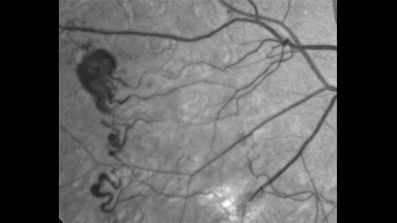
Along with their cross-sectional images, commercially available OCT scanners produce near-infrared images that show hyporeflective foci. These foci indicate hemorrhages that are characteristic of diabetic retinopathy.
“That’s actually how we rate diabetic retinopathy severity in general,” Naguib said. “We basically count the number of hemorrhages that we see. And, depending on the number of hemorrhages, we determine whether a person has mild or moderate or severe retinopathy. This is using the same thing except instead of seeing it in red, you’re basically seeing it in this kind of dark shade.”
The system isn’t perfect because other aspects of the retina can appear as dark shades, but it might be good enough for screening purposes, he added.
To put the approach to the test, the researchers reviewed charts to identify patients with type 1 and type 2 diabetes with and without diabetic retinopathy.
They randomly sampled these charts to generate matched cohorts of patients with all stages of diabetic retinopathy. They excluded patients with low-quality OCT scores or other macular diseases. And they only included hyporeflective foci with discrete margins to avoid mislabeling of signals from retinal pigment epithelium changes.
They identified 195 patients who were then roughly evenly divided into groups of having no diabetic retinopathy, mild, moderate, severe, or proliferative disease.
Two clinicians who didn’t know the patients’ diagnoses counted the number of hyopreflective foci in each infrared image. A third clinician resolved any discrepancies of more than 10 hyporeflective foci.
The mean number of hyporeflective foci for patients without diabetic retinopathy was 1.9. For mild disease it was 4.6; for moderate disease, 10.4; for severe disease, 12.2; and for proliferative disease, 12.1.
The differences were statistically significant for no disease on one hand and moderate, severe, or proliferative disease on the other (P < .0001).
They were also significant for the differences between mild and moderate disease (P < .008); and between moderate and either severe or proliferative disease (P < .0001).
The area under the receiver operator curve was 0.849 (95% CI, 0.792-0.905). Based on this curve, the researchers determined that the ideal threshold for detection of moderate nonproliferative diabetic retinopathy or worse was 4.75 hyporeflective foci, with a sensitivity of 80%.
“Obviously, it could be better,” said Naguib. “But in this small study, we felt like that was fairly compelling.”
The next step in the research would be a more rigorous study comparing the near- infrared images with standardized photos that were graded in a standard way, he said.
The researchers are also exploring other vascular abnormalities in the infrared images, in hope that they can use them to differentiate among the advanced stages of diabetic retinopathy. In the future, they also hope to use artificial intelligence to increase accuracy and practicality of the screening.
The approach looks promising if some way can be found to avoid the high cost of OCT machines, said session co-moderator Jennifer Lim, chair of ophthalmology at the University of Illinois Hospital & Health Sciences System in Chicago. “Could you get a near-infrared device that’s not as expensive as OCT?” she asked.
The cutoff between mild and moderate disease seems practical because it is similar to what is used with other diagnostic tools, she told Medscape Medical News. However, she wondered if the near infrared images generated by OCT have a wide enough field to capture all the hyopreflective foci that would be useful in screening for diabetic retinopathy.
“If we relegated just to the immediate macular region, we’re going to miss more than we would want, I think,” she said.
Naguib reports no relevant financial relationships. Lim reports relationships with Alcon, Aldeyra, Allergan, Aura, Chengdu Pharmaceuticals, Cognition, Eyenuk, Genentech/ Roche, Graybug, Iveric Bio, Luxa, NGM, Novartis, Opthea, Quark, R egeneron, RGM, Santen, Stealth, Unity, and Veridian.
American Society of Retina Specialists (ASRS). Presented July 14, 2022
Laird Harrison writes about science, health and culture. His work has appeared in national magazines, in newspapers, on public radio, and on websites. He is at work on a novel about alternate realities in physics. Harrison teaches writing at the Writers Grotto. Visit him at www.lairdharrison.com or follow him on Twitter: @LairdH.
Follow Medscape on Facebook, Twitter, Instagram, and YouTube
Source: Read Full Article


