Clathrin-mediated Endocytosis
In clathrin-mediated endocytosis, the cell membrane invaginates to take up metabolites, hormones, and proteins from the cell surface into intracellular vesicles.
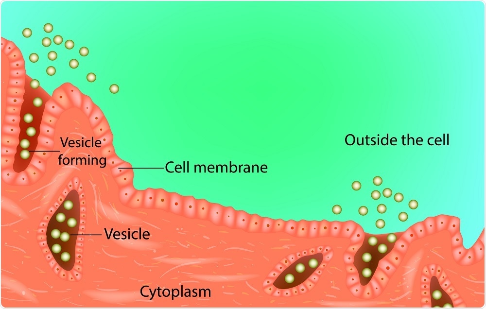
Sakurra | Shutterstock
Cells employ several pathways for protein and lipid intake from the cell surface. These pathways include micropinocytosis, phagocytosis, and clathrin-mediated or caveolin-mediated endocytosis.
Clathrin-mediated endocytosis is involved in turnover of plasma membrane proteins, uptake of low-density lipoproteins, and endocytosis of several growth factors. Many viruses, such as influenza and shigella, may also hijack clathrin-mediated endocytosis to gain cell entry.
Initiation of clathrin-coated pit (CCP) formation
The clathrin coat is comprised of triskelion, a molecule which consists of three copies of clathrin-heavy chains linked by trimeric domains.
The CCP can be divided into several segments: proximal leg, distal leg, ankle, and knee. In neuronal tissues, each clathrin heavy chain is associated with two clathrin light chains in a 1:1 ratio.
The clathrin light chain can disrupt contact with the clathrin heavy chain to prevent unwanted assembly in the cytoplasm. Furthermore, electron microscopy of clathrin-coated vesicles shows that clathrin light chains are positioned near the cytosol. This positioning may improve its interaction with regulatory elements present in the cytoplasm.
Role of clathrin and AP-2
Initially, triskelion proteins form a scaffold and recruit several clathrin-associated proteins on the cell membrane. As triskelion itself does not bind to the membrane, it requires factors to stabilize its association with the membrane.
One such adaptor is AP-2, which consists of α, β2, μ2, and σ2 adaptin subunits. When AP-2 is depleted, the clathrin-membrane association also decreases; thus, AP-2 appears to have a role in clathrin recruitment.
PIP2 binds to AP-2 subunits and plays a role in its recruitment. PIP2 formation is regulated by lipid kinases that convert PIP to PIP2.
Nucleation sites for CCPs
An important question in clathrin-mediated endocytosis is whether CCPs arise in predetermined or stochastic sites. In the presynaptic nerve terminal, there appear to be hot spots where pits emerge at higher rates.
One scaffold protein in these membrane hotspots is the 160-kDa dynamin-associated protein (DAP160). In Drosophila, DAP160 surrounds the active zone and contains AP-2. In cases where DAP160 function is abrogated, the synaptic vesicles fail to form.
Phosphorylation
Several kinases and phosphatases are involved in the regulation of clathrin-mediated endocytosis. Phosphorylation may serve as a positive or negative regulator. For example, adaptor-associated kinase 1 (AAK1) can phosphorylate the μ2 subunit of AP-2, which then undergoes a conformational change and binds to PIP2 and other cargo binding proteins.
Similarly, tyrosine kinase has been shown to phosphorylate clathrin heavy chains and positively regulate clathrin-mediated endocytosis. Casein kinase II (CKII) is an example of a kinase that negatively regulates clathrin-mediated endocytosis via phosphorylation. Calcineurin, a Ca2+-activated protein phosphatase, also regulates synaptic vesicles.
Membrane curvature
Following nucleation, the membrane changes conformation from a flat bilayer to develop curvature. One method by which curvature is achieved is through the use amphipathic helices to create asymmetry in the bilayer.
COPII vesicles and BAR domains are also involved in inducing membrane curvature. The BAR domain is comprised of a positively-charged, elongated crescent across a concave surface. The positive charge leads to electrostatic interactions with the membrane that induce curvature.
Scission
Dynamin is a critical molecule involved in vesicle budding. It consists of a GTPase domain, PH domain, GTPase effector domain (GED), and proline-rich domain (PRD). The PH domain binds to PIP2 , whereas the PRD domain binds to SH3 domain proteins. Dynamin uses GTP hydrolysis to generate a twisting force which leads to vesicle scission.
Sources:
- https://www.ncbi.nlm.nih.gov/books/NBK6479/
- www.mechanobio.info/…/
- www.annualreviews.org/doi/abs/10.1146/annurev-biochem-062917-012644
Further Reading
- All Cell Signaling Content
- The cAMP signaling pathway
- Phosphatidylinositol Biphosphate (PIP2) Signal Pathway
- GPCR Signaling Pathways
- GPCR Origin and Diversification
Last Updated: Dec 3, 2018

Written by
Dr. Surat P
Dr. Surat graduated with a Ph.D. in Cell Biology and Mechanobiology from the Tata Institute of Fundamental Research (Mumbai, India) in 2016. Prior to her Ph.D., Surat studied for a Bachelor of Science (B.Sc.) degree in Zoology, during which she was the recipient of anIndian Academy of SciencesSummer Fellowship to study the proteins involved in AIDs. She produces feature articles on a wide range of topics, such as medical ethics, data manipulation, pseudoscience and superstition, education, and human evolution. She is passionate about science communication and writes articles covering all areas of the life sciences.
Source: Read Full Article