Studying the human brain in mice

The human brain is a tricky study subject. Brain scans are still limited in resolution and the knowledge they can provide, and in vitro approaches are not yet able to fully replicate the important micro-environment of brain cells. A new method developed by the lab of Bart De Strooper (VIB-KU Leuven Center for Brain & Disease Research) pioneers the transplantation of human microglia cells into mice brains. Their work appears in Nature Neuroscience.
Microglia & Alzheimer’s
Microglia cells, brain cells that are responsible for brain ‘maintenance,” are thought to play an important role in the development of Alzheimer’s disease. But they are not easy to study.
Culturing them in petri dish neglects the complex environment in which they usually function, and model organism, such as mice, have microglia that are too different from human ones to draw robust conclusions.
Human cells in a mouse brain
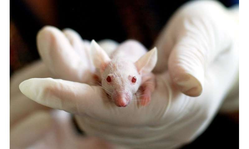
Now, the lab of Bart De Strooper (VIB-KU Leuven Center for Brain & Disease Research) managed to derive human microglia from stem cells and transplant them into the brains of mice.
Prof. De Strooper explains: “This paper is the first to investigate how human microglia react in Alzheimer’s disease. By bringing living human microglia in the mouse brain we were able to see how they attack the human amyloid peptide. This work allows us to investigate how a combination of human genes that increase risk of Alzheimer’s disease work in human brain cells.”
Dr. Renzo Mancuso, the first author of the paper, adds: “The remarkable resemblance between the human microglia transplanted in the mouse brain and the human microglia isolated from living patients that were undergoing neurosurgery was spectacular and demonstrated that this work is extraordinary relevant for human biology.”
Source: Read Full Article


