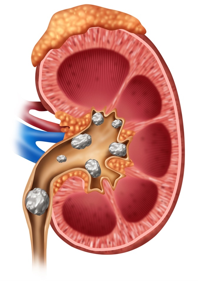
Kidney Stone Diagnosis
To diagnose kidney stones physicians ask patients about the symptoms and health conditions that could possibly promote stone formation in the kidneys, and review the patient’s medical history, family history of stone formation, and foods consumed. Laboratory and imaging tests are carried out after physical tests to diagnose the presence of kidney stones.

Biochemistry Tests
The acidity of the urine is measured by a pH test. The normal range of urine pH is 4.6– 8.0. When the value is below 5.5, there is a risk of developing uric acid stones. When the value is above pH 6.2, there is a higher chance of calcium phosphate stones.
We can infer uric acid levels or calcium levels in the blood from the blood test results. They also help in monitoring the kidney health. Some physicians also advise antacid along with meals for reducing the acid concentration in the urine.
The urine test reveals the mineral levels—whether too low to form stones or too high to potentially form kidney stones. The risk of developing calcium stones is high when the urine has more than 200 mg of calcium/24 h.
Imaging Tests
Physicians carry out imaging tests to locate kidney stones. The imaging tests show the issues leading to stone formation, that is, whether it is a birth defect or a block in the urinary tract. Anesthesia is not used for imaging tests.
Abdominal X-Rays
Abdominal X-rays use low radiation levels and capture pictures of the abdomen. The X-rays are recorded on a computer screen or on a photographic film. A radiologist analyzes the images and provides his comments. Though abdominal X-rays do not display all stones, they can pinpoint the exact location of a kidney stone.
Intravenous Pyelogram (IVP)
IVP, an X-ray test, makes use of a special substance that outlines the bladder, ureters, and kidneys and helps see the manner in which the urinary system is handling waste products.
By using IVP, physicians can find out issues present in the urinary tract. As IVP can display the shape of the kidneys and the components of the urinary system, it is used to find an enlarged prostate, kidney stones, urinary tract defects, scarring caused by infection in the urinary tract, and tumors of the urinary system.
The dye used in the tests helps to see the organs clearly in the X-ray. IVP is also used to find out the reason for blood in the urine or pain in the lower/side back, which are both generally symptoms of kidney stones.
With the advent of computed tomography (CT), IVP is no longer widely used, although they expose a patient to less radiation than CT scans do.
Retrograde Pyelography
Similar to IVP, retrograde pyelography is used to obtain a clear picture of the upper urinary tract comprising the kidneys and ureters. This test belongs to the X-ray category, where a contrast agent is used to view the kidneys or ureters clearly. Unlike IVP where the contrast agent is injected into the vein, in retrograde pyelography, the contrast agent is injected into the ureter. Even though newer diagnostic tests that have replaced retrograde pyelography are in use, the details of the upper urinary tract can be viewed better with retrograde pyelography. When IVP is not found to be effective, retrograde pyelography is suggested.
Kidney Ultrasound
Kidney ultrasound—a noninvasive test—produces images using the shape, size, and location of the kidneys as well as the structures of bladder and ureters and the flow of blood to the kidneys can be examined.
The ultrasound is used to find out the amount of fluid that is collected, and cysts, tumors, stones, and obstructions and infections in or surrounding the kidneys and ureters. The procedure does not involve any radiation and causes no discomfort to the patients.
Abdominal MRI (Magnetic Resonance Imaging)
When physicians suspect kidney stones, they prefer the patients to undergo an abdominal MRI scan. The imaging test does not use X-rays; instead it makes use of radio waves and magnetic waves that are powerful enough to create pictures of the interior portion of the abdomen. Single MRI images known as slices can be saved on a computer or a photographic film. Numerous images can be produced in a single test.
Computed Tomography (CT)
The image of the urinary tract is created with the help of computer technology and X-rays. Generally, a contrast medium is not used in a CT scan to see the urinary tract; however, physicians may suggest using a contrast medium to clearly visualize the parts that comprise the urinary tract. Patients are asked to lie down on a table that moves through a device resembling a tunnel so that X-rays can be taken. CT scans also help locate the position and size of the stones.
Sources
- www.niddk.nih.gov/…/diagnosis
- www.mayoclinic.org/…/dxc-20319568
- www.urologyhealth.org/urologic-conditions/intravenous-pyelogram-(ivp)
- www.urologyhealth.org/urologic-conditions/retrograde-pyelography
- www.hopkinsmedicine.org/…/
- https://medlineplus.gov/ency/article/003796.htm
- www.kidney.org/…/11-10-1815_HBE_PatBro_Urinalysis_v6.pdf
- www.pathology.leedsth.nhs.uk/…/RenalStones.aspx
Further Reading
- All Kidney Stone Content
- Diet and Nutrition for Kidney Stones
- Kidney Stones: Symptoms and Causes
- Kidney Stones – Nephrolithiasis
- Kidney Stones – Treatment
Last Updated: Feb 26, 2019
Source: Read Full Article


