Small glowing protein allows researchers to peer deeper into living tissues: Proteins that emit longer wavelengths of near-infrared light help create detailed, hi-res biomedical images
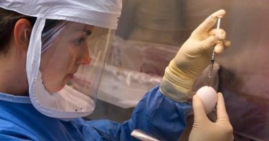
Biomedical and genetic engineers at Duke University and the Albert Einstein College of Medicine have designed a small fluorescent protein that emits and absorbs light that penetrates deep into biological tissue. Tailored to wavelengths in the near-infrared (NIR) spectrum, this protein can help researchers capture deeper, cleaner, more precise biomedical images.
This work appeared Dec. 1 in the journal Nature Methods.
Imaging deep tissues with light is challenging. Visible light is often quickly absorbed and scattered by structures and molecules in the body, preventing researchers from seeing deeper than a millimeter within a tissue. If they do manage to probe further, substances like collagen or melanin often muddy the image, creating the equivalent of background noise through their natural fluorescence.
“Biological molecules naturally absorb and emit light in the visible spectrum, which is about 350 to 700 nanometers,” said Junjie Yao, assistant professor of biomedical engineering at Duke. “So when using it to image deep tissue, it’s like trying to observe the stars in daylight. The signals get flooded out.”
To wade out from these muddied waters, Yao and his collaborator Vladislav Verkhusha, professor of genetics at Albert Einstein College of Medicine, New York developed a protein that absorbs and emits longer wavelengths of light in the near-infrared (NIR) spectrum.
“Tissue is the most transparent in the 700-1300 nanometer window of NIR light,” said Yao. “At those wavelengths, light can penetrate deeper into a tissue, and because there is less natural background fluorescence to filter out, we can take longer exposures and capture clearer images.”
Verkhusha and his lab used a process called directed molecular evolution to engineer their proteins, using photoreceptors normally found in bacteria as the basis for the structure. These photoreceptors are useful for imaging research because they can switch between a silent and active state when hit with a specific wavelength of light. They can bind with biliverdin, a biomolecule that appears in high quantities in mammalian and human tissues.
Source: Read Full Article


