Researchers target proteins, pathways behind congenital heart disease

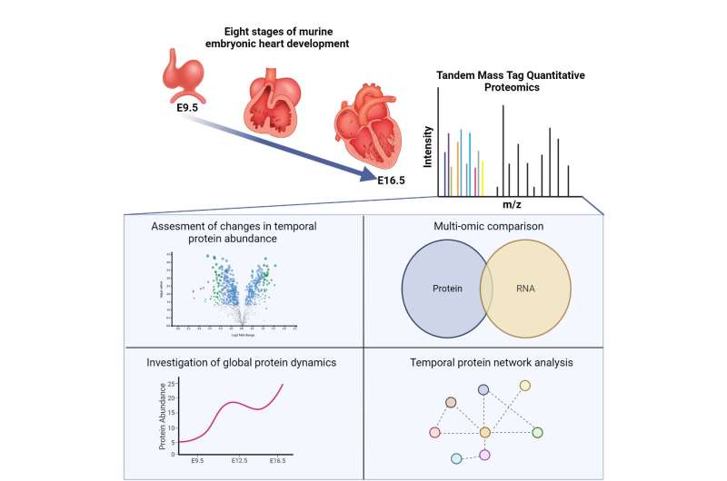
Researchers at the UNC School of Medicine, UNC McAllister Heart Institute, and the UNC Lineberger Comprehensive Cancer Center characterized the expression of thousands of cardiac proteins during eight critical stages of embryonic heart development.
This research, published in Development Cell, will provide scientists with much-needed information to identify biological causes for congenital heart disease, or CHD.
“We now have a foundational data set that shows how protein dynamics change in normal heart development,” said first author Whitney Edwards, Ph.D., and assistant professor in the UNC Department of Cell Biology and Physiology. “Researchers can use this as the blueprint to figure out the specific pathways or proteins contributing to congenital heart disease.”
The formation of the human heart
The development of the human heart is a very complex process. Congenital heart disease (CHD), one of the most prevalent congenital diseases, occurs when a person is born with one or more structural flaws in the heart or its larger vessels. While some types of CHD are mild, other more complex defects cause life-threatening complications.
Historically, research into what causes normal and abnormal heart development has mainly focused on genomics and gene expression. But much less effort has been given to directly assessing cardiac proteins. While genes contain the instructions needed to make proteins, many factors can lead to discrepancies between gene and protein expression.
To understand the underlying processes of heart development and CHDs, it is critical to study both the genome and proteome, which is the set of expressed proteins in a given type of cell or organism.
Measuring protein dynamics
In this study, Edwards and colleagues took advantage of a technique known as multiplexed quantitative proteomics, an innovative protein labeling technique that allows them to identify and measure the abundance of a large number of proteins from relatively small amounts of heart tissue.
Using this method, they measured the expression of 7,313 cardiac proteins and determined that approximately 3,799 proteins show differences in expression over the course of heart development. Their findings can now be used to determine when particular proteins and molecular pathways are active during specific stages of heart development.
The mevalonate pathway
Surprisingly, analysis of the cardiac proteome revealed that proteins in the mevalonate pathway, an essential metabolic pathway, are highly expressed midway through heart development. This pathway produces many important biomolecules, such as cholesterol, isoprenoids, and vitamin K.
Through their thorough analysis, the researchers determined that the mevalonate pathway regulates embryonic heart cell cycling and critical signaling molecules. They now believe that the pathway controls the cell’s ability to divide and, therefore, may be essential for the overall growth of the developing heart.
Edwards and colleagues also found that the mevalonate pathway has close ties with a particular signaling protein called Yap, which is incredibly important in the function and regulation of cardiac development, homeostasis, and regeneration.
The Edwards’ lab is now focused on combining their proteomics data set with human genetics data to identify novel targets causing CHD.
More information:
Whitney Edwards et al, Quantitative proteomic profiling identifies global protein network dynamics in murine embryonic heart development, Developmental Cell (2023). DOI: 10.1016/j.devcel.2023.04.011
Journal information:
Developmental Cell
Source: Read Full Article


