Mycobacterium tuberculosis may induce resistance to secondary SARS-CoV-2 infection
A team of United States-based scientists recently conducted a study to evaluate the effect of Mycobacterium tuberculosis infection on the pathogenesis of severe acute respiratory syndrome coronavirus 2 (SARS-CoV-2) infection.
The findings of this study, which is published on the preprint server bioRxiv*, reveal that mice infected with Mycobacterium tuberculosis are resistant to SARS-CoV-2 infection. This resistance is induced by expansion of T- and B-cell subsets in the lungs upon secondary viral challenge.
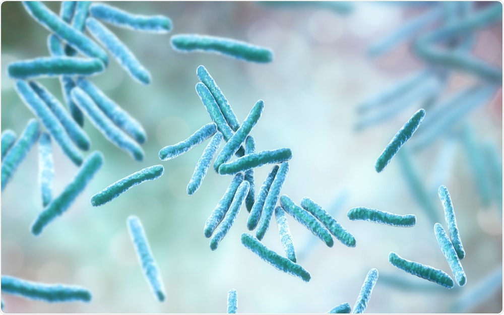
 Study: Mice infected with Mycobacterium tuberculosis are resistant to secondary infection with SARS-CoV-2. Image Credit: Kateryna Kon / Shutterstock.com
Background
Mycobacterium tuberculosis is a pathogenic bacterium belonging to the family Mycobacteriaceae. Like SARS-CoV-2, the causative pathogen of coronavirus disease 2019 (COVID-19), Mycobacterium tuberculosis causes severe and often fatal lung infection called tuberculosis.
Both tuberculosis and COVID-19 are associated with high mortality rates in humans. Interestingly, there is evidence suggesting that the mortality rate of COVID-19 is relatively low in countries where tuberculosis is prevalent.
In the current study, the scientists investigate the clinical consequences of mycobacterium tuberculosis and SARS-CoV-2 co-infection in mice.
About the study
The scientists developed two mouse models of COVID-19 using mice that were chronically infected with Mycobacterium tuberculosis. To this end, human angiotensin-converting enzyme 2 (ACE-2)-expressing mice were infected with low-dose mycobacterium tuberculosis via aerosol-based delivery.
After 30 days, secondary SARS-CoV-2 infection was induced in these mice through the intranasal route. The researchers subsequently assessed the clinical consequences of co-infection at days 4, 7, and 14 post-viral challenge. The controls in this study were mice infected with either Mycobacterium tuberculosis or SARS-CoV-2.
Clinical consequences of co-infection
The highest reduction in body weight was observed in mice infected with only SARS-CoV-2. Interestingly, co-infected mice did not show any significant body weight loss and were comparable to mice infected with only Mycobacterium tuberculosis.
A significantly lower lung viral load was observed in co-infected mice compared to SARS-CoV-2-infected mice. Moreover, no change in the growth of Mycobacterium tuberculosis was observed in the lungs, liver, and spleen after the viral challenge.
Immune response to co-infection
The challenge of mice with SARS-CoV-2 caused a significant increase in the levels of proinflammatory mediators including interferon g (IFN-g), interleukin-6 (IL-6) and IL-1b. Mice infected with Mycobacterium tuberculosis only exhibited even higher levels of inflammatory mediators in the lungs.
Importantly, the levels remained unchanged upon challenge with SARS-CoV-2. The resistance of Mycobacterium tuberculosis-infected mice to SARS-CoV-2 was not associated with an elevated expression of anti-inflammatory mediators.
Regarding histopathological changes, SARS-CoV-2-infected mice showed significant levels of alveolar necrosis and infiltration of proinflammatory mediators. A significantly higher level of pneumonia and hyaline membrane formation was observed in the lungs of SARS-CoV-2-infected mice. However, these changes were not seen in the lungs of co-infected mice.
The viral resistance due to Mycobacterium tuberculosis infection observed in ACE-2-expressing mice was also observed in mice infected with mouse-adapted SARS-CoV-2. Immune cells isolated from the lungs of mice infected with mouse-adapted SARS-CoV-2, Mycobacterium tuberculosis, or both were subjected to single-cell ribonucleic acid (RNA) sequencing to determine the mechanism of Mycobacterium tuberculosis-induced viral resistance.
The findings of RNA sequencing revealed that the immune environment in the lungs of co-infected mice was similar to that observed in the lungs of Mycobacterium tuberculosis-infected mice, with the exception of expanded B-cell and T-cell subsets.
Study significance
The current study reveals that mice infected with Mycobacterium tuberculosis are resistant to secondary SARS-CoV-2 infection and COVID-19-related pathologies. To this end, the Mycobacterium tuberculosis infection appears to create an inflammatory microenvironment in the lungs, which is unfavorable for SARS-CoV-2 propagation.
The presence of a wide variety of innate immune cells due to Mycobacterium tuberculosis infection may prevent SARS-CoV-2 infection. In addition, Mycobacterium tuberculosis-induced adaptive immune response may cross-react with viral antigen to induce resistance. The expansion of B-cell and T-cell subsets after the SARS-CoV-2 challenge supports the explanation of Mycobacterium tuberculosis-induced SARS-CoV-2 resistance.
*Important notice
bioRxiv publishes preliminary scientific reports that are not peer-reviewed and, therefore, should not be regarded as conclusive, guide clinical practice/health-related behavior, or treated as established information.
- Mejia, O.R., Gloag, E. S., Ruane-Foster, M., et al. (2021). Mice infected with Mycobacterium tuberculosis are resistant to secondary infection with SARS-CoV-2. bioRxiv. doi:10.1101/2021-11.09.467862. https://www.biorxiv.org/content/10.1101/2021.11.09.467862v1
Posted in: Medical Science News | Medical Research News | Disease/Infection News
Tags: Angiotensin, Angiotensin-Converting Enzyme 2, Antigen, Anti-Inflammatory, Cell, Coronavirus, Coronavirus Disease COVID-19, Enzyme, Immune Response, Interferon, Interleukin, Interleukin-6, Liver, Lungs, Membrane, Mortality, Necrosis, Pathogen, Pneumonia, Propagation, Respiratory, Ribonucleic Acid, RNA, RNA Sequencing, SARS, SARS-CoV-2, Severe Acute Respiratory, Severe Acute Respiratory Syndrome, Spleen, Syndrome, T-Cell, Tuberculosis, Weight Loss

Written by
Dr. Sanchari Sinha Dutta
Dr. Sanchari Sinha Dutta is a science communicator who believes in spreading the power of science in every corner of the world. She has a Bachelor of Science (B.Sc.) degree and a Master's of Science (M.Sc.) in biology and human physiology. Following her Master's degree, Sanchari went on to study a Ph.D. in human physiology. She has authored more than 10 original research articles, all of which have been published in world renowned international journals.
Source: Read Full Article


