Elevated and sustained autoantibodies following COVID-19
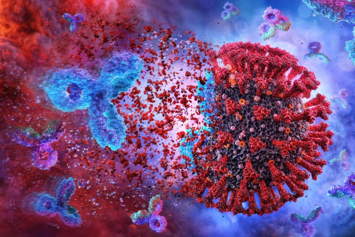
A recent study posted to the medRxiv* preprint server demonstrated that autoantibody levels rise following acute severe acute respiratory syndrome coronavirus 2 (SARS-CoV-2) infection and remain to be elevated for about six months.

Background
Existing reports suggested that people hospitalized for SARS-CoV-2 infection had high autoantibody concentrations reactive to self-epitopes such as interferons and phospholipids with a possible functional significance. Prior studies have documented links between autoantibody levels and SARS-CoV-2 severity.
However, temporal patterns and amounts of these autoantibodies months after CoV disease 2019 (COVID-19) remain unknown. Indeed, it is unclear if recovered SARS-CoV-2-infected people's composite autoantibody profiles are similar to those with confirmed autoimmune disorders.
About the study
The present study aimed to discover more about the relationships between SARS-CoV-2 infection and autoimmunity. The scientists assessed the circulatory concentrations of 17 autoantibodies correlated to autoimmune connective tissue disorders from COVID-19 inpatient and outpatient participants. They also analyzed these autoantibody levels in people with systemic lupus erythematosus (SLE), non-infected pre-pandemic controls, and scleroderma (SSc).
The SARS-CoV-2-infected outpatient and hospitalized subjects represented those with mild and severe COVID-19, respectively. The researchers contrasted autoantibody concentrations from both late and early timestamps (≥90 or ≤30 days after symptom onset) of the hospitalized and outpatient COVID-19 patients with two autoimmune participant cohorts (SSc and SLE) and uninfected pre-pandemic controls. They performed multivariate analyses to evaluate the mechanism by which COVID-19 correlated with autoantibody positivity considering the co-morbidities and demographics of the participants.
In addition, the researchers examined the longitudinal trajectory of autoantibodies months following COVID-19 symptom onset. For this, the team assessed various trends of change in personal autoantibody concentrations in all SARS-CoV-2-infected subjects over time (two to five timestamps per individual, extending about six months). They also used partial least squares discriminant analysis (PLS-DA) to examine the individual autoantibody expression fingerprints of COVID-19 patients months after convalescence relative to those with autoimmune disorders and uninfected controls.
Results
The study results demonstrated that seven out of the 17 autoantibodies tested were elevated in inpatient or outpatient SARS-CoV-2 patients nearly six months following symptom start than controls. The seven autoantibodies were anti-alanyl-transfer ribonucleic acid (tRNA) synthetase (PL-12), Ku, anti-topoisomerase (Scl-70), β-2-glycoprotein, proteinase 3, ribonucleoprotein (RNP)/anti-Smith (Sm), and sjögren syndrome type B (SSB/La). Furthermore, multivariate analyses revealed associations between COVID-19 and the positivity of SSB/La, Sm, myeloperoxidase, proteinase 3, histidyl-tRNA synthetase 1 (Jo-1), and Ku reactive immunoglobulin Gs (IgGs) six months after symptom onset.
COVID-19 patients' autoantibody levels were tracked for about six months from the beginning of symptoms, and various temporal autoantibody patterns were identified. SARS-CoV-2-infected subjects possessed a greater autoantibody level than unexposed controls at both convalescent and acute timestamps. Autoantibody expression profiles of each participant displayed similarities between recovered SARS-CoV-2-infected and pre-pandemic groups, which were unique from the SLE and SSc subjects. These findings suggest that COVID-19 convalescents experience a random, disorganized autoantibody generation, bolstering the proposed processes, like lymphopenia-induced loss of tolerance, different from epitope spreading or molecular mimicry.
In 18% of outpatients and 53% of inpatient participants, a negative, then positive expression trend for at least one autoantibody was discovered, showing the induction and long-term expression of self-reactive immune responses after COVID-19, especially in severe acute disease. Furthermore, the positivity of a major autoantibody temporal expression profile in COVID-19 outpatient and inpatient patients at all timestamps indicated an extremely early generation of autoantibody or its pre-existence before viral exposure.
Conclusions
According to study findings, autoantibodies connected to autoimmune connective tissue pathologies were found in COVID-19 patients months after convalescence than in pre-pandemic controls. Further, the investigation showed temporal pathways denoting the commencement of novel autoimmune responses following SARS-CoV-2 infection.
The study findings suggested that SARS-CoV-2 leaves a significant autoimmune imprint at least in the initial six months following infection. Unlike participant samples collected before the COVID-19 pandemic, autoantibodies in convalescent SARS-CoV-2-infected subjects were significantly greater. Even when the SARS-CoV-2 infection occurred about six months earlier, COVID-19 history showed significant correlations with the presence of numerous autoantibodies after controlling for participant medical conditions and demographics.
The authors mentioned that because autoantibody positivity can arise years before the onset of autoimmune illness, the idea that SARS-CoV-2-linked autoantibodies represent a precursor to future autoimmune diseases warrants additional examination. Moreover, understanding the interaction between variables, like immune memory and pre-existing infections, novel-onset immune responses, acute viral infection and inflammation, and lymphopenia, was critical to combat SARS-CoV-2-linked morbidity and death.
*Important notice
medRxiv publishes preliminary scientific reports that are not peer-reviewed and, therefore, should not be regarded as conclusive, guide clinical practice/health-related behavior, or treated as established information.
- Nahid Bhadelia, Alex Olson, Erika Smith, Katherine Reifler, Jacob Cabrejas, Maria Jose Ayuso, Katherine Clarke, Rachel Ruby Yuen, Nina Lin, Zach Manickas-Hill, Ian Rifkin, Andreea Monica Bujor, Manish Sagar, Anna Belkina, Jennifer Snyder-Cappione. (2022). Longitudinal analysis reveals elevation then sustained higher expression of autoantibodies for six months after SARS-CoV-2 infection. medRxiv. doi: https://doi.org/10.1101/2022.05.04.22274681 https://www.medrxiv.org/content/10.1101/2022.05.04.22274681v1
Posted in: Medical Science News | Medical Research News | Disease/Infection News
Tags: Autoantibodies, Autoimmunity, Coronavirus, Coronavirus Disease COVID-19, covid-19, Glycoprotein, Immunoglobulin, Inflammation, Interferons, Lupus, Lupus Erythematosus, Lymphopenia, Myeloperoxidase, Pandemic, Respiratory, Ribonucleic Acid, SARS, SARS-CoV-2, Scleroderma, Severe Acute Respiratory, Severe Acute Respiratory Syndrome, Syndrome, Systemic Lupus Erythematosus

Written by
Shanet Susan Alex
Shanet Susan Alex, a medical writer, based in Kerala, India, is a Doctor of Pharmacy graduate from Kerala University of Health Sciences. Her academic background is in clinical pharmacy and research, and she is passionate about medical writing. Shanet has published papers in the International Journal of Medical Science and Current Research (IJMSCR), the International Journal of Pharmacy (IJP), and the International Journal of Medical Science and Applied Research (IJMSAR). Apart from work, she enjoys listening to music and watching movies.
Source: Read Full Article


