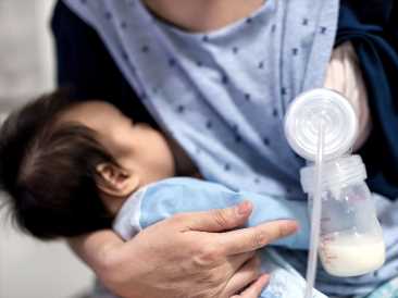
Breast cancer: Alive cells from human breast milk could offer insights

- Medical researchers have assumed that breast cells found in breast milk were dead or dying, but a recent study shows these cells are alive and can be isolated and studied.
- Genetic analysis of these cells has revealed two cell populations responsible for secreting breast milk in human breasts, whereas previously there was assumed to be just one as observed in mice.
- Future studies of these cells could provide insight into how breast cancer originates and how breastfeeding could protect some individuals.
The protective effect of breastfeeding against breast cancer has been known for some time. However, the mechanisms underlying this have remained unclear.
While mouse studies suggest this could be due to changes in the breast tissue that occur during and breastfeeding causing long lasting epigenetic changes, it has been difficult to investigate. This is because most breast tissue donors are people undergoing surgery and very few of these individuals would have been lactating.
During breastfeeding, some of the cells from the surface of the milk glands inside the breast are expelled into the breast milk itself. While it was not clear how much insight they could provide as scientists had previously assumed them to be dead or dying, a recent study by a team from the Wellcome-MRC Cambridge Stem Cell Institute (CSCI) and the Department of Pharmacology at the University of Cambridge, UK, has shown that these remain alive in the milk and can be isolated from expressed breast milk.
In a paper in Nature Communications, the team outlined their findings by analyzing these breast cells for the first time.
Karis Betts, health information manager at Cancer Research UK — who was not involved in the research — told Medical News Today in an email:
“Current research suggests a link between breastfeeding and reduced risk of breast cancer. Not all questions have been answered about why this is and how it affects different groups of women and different types of breast cancer. It’s thought that hormonal changes, impact on ovulation, and changes to the cells in the breast might play a role.”
“This early-stage research shows the difference between cell types present when lactating and not, which helps paint the picture of what these cell changes might be. But the study can’t tell us which of these changes, if any, are linked to breast cancer in people.”
Epigenetic changes
To determine how the genes that are activated in breastfeeding women differ from those not breastfeeding, researchers isolated breast cells from milk donated from nine women who had been breastfeeding for less than a year and carried out a genetic analysis. They compared these to cells from tissue donated by seven women having aesthetic breast reduction surgery.
A form of genetic analysis called single-cell RNA sequencing showed genes that increase their expression during lactation related to fatty acid metabolism and storage, zinc transport, and secretion and immune response.
They also identified two distinct cell populations responsible for making breast milk in lactating women.
This differs from previous research showing just one type of cell in mice. Researchers were surprised to find that one of these groups of cells had high levels of expression of genes responsible for immune response not previously associated with human breast cells.
This finding alone reframed the question of why breast cells were found in human milk at all. Researchers suggested they could serve a role in helping the breastfed infant’s immune system be ‘trained’.
“I believe that by studying human milk cells, we will be able to answer some of the most fundamental questions around mammary gland function such as: how is milk produced? Why do some women struggle to make milk? and what strategies can be employed to improve breastfeeding outcomes for women?” says Dr. Alecia-Jane Twigger from the Wellcome-MRC Cambridge Stem Cell Institute, who led the study.
Insight into breast cancer origins
Researchers then performed further analyses to determine the origin of these cells, and researchers concluded they had differentiated from luminal progenitor cells as they had similar genetic profiles. Researchers concluded that determining the exact differentiation pathway would involve sampling cells or tissues as they differentiated in pregnant women, as lactation starts during pregnancy and then production increases when the placenta detaches from the womb after childbirth.
Luminal progenitor cells are significant to researchers as they are thought to be the cells in which cancerous mutations first originate.
Lead author Dr. Walid Khaled from the Wellcome-MRC Cambridge Stem Cell Institute said: “We’re not saying that the cells [found in breastmilk] themselves are going to go and give rise to cancer because that’s not true […] But because they do resemble the population of cells known as luminal progenitors, and probably are daughters of luminal progenitors, and have characteristics of luminal progenitors, that all of a sudden enables us to do studies on these cells with a lot of ease.”
Accessing donated breast milk had proved easy as many donors were willing, he said. Though the number of women contributing to this study was small, Dr. Khaled suggested that looking at changes in the breast following subsequent pregnancies using cells isolated from breastmilk could provide further insight into women’s health.
“You could imagine collecting a milk sample from a first pregnancy, and then once every now and then down the line from a second or third pregnancy. Then all of a sudden, you’ve got a window on how the breast has changed over that time, right? So I think that could potentially be really exciting.”
Source: Read Full Article


